
Arachnoidiscus sendaicus
 This Arachnoidisucus sendiacus is 453 microns or 0.453 mm: a large diatom. Diatoms are single celled algae with a glass cell wall.
The image is stitched together from about thirty images, each of which where composites of two images at different depths of field. The original magnification was x1000, using the most powerful type of microscope lens. High power lenses require immersion in an oil with a high refractive index to cause the light rays to spread out to increase resolution. The tiny blobs, called punctae, in the centre are about one micron. Thats just two wavelengths of visible light, and close to the limit of resolution of a light based microscope.
This photograph was constructed from a slide I bought from perhaps the most famous slide maker and diatomist, Klaus Kemp. Every other image I have made is from samples/slides I have prepared.
Shades of Grey

A penntate diatom, again from a slide made by Klaus Kemp. Sadly I don’t have an id on this one, but will update when I do. This is 198 microns wide. I used a yellow filter, which slightly improves the resolution of the x100 oil immersion objective lens, x1000 with the second photo-relay lens, and then shot only in shades of grey.
Lots of little dots

Thalassiosira baltica a marine diatom 103 microns again many thanks to Klaus, again using the immersion lens at x1000. I have inverted the colours digitally. The image seems even more striking in this form.
Neon

Triceratium nobolis a marine diatom 93 microns from 102 form slide by Klaus Kemp, again using the immersion lens at x1000. I have inverted the colours digitally. A ghostly triangle of mercury gas discharge tube colours. As discharge tubes are commonly referred to as Neon lights I gave this simple name.
Coscinodiscus robustus

Coscinodiscus robustus A marine diatom 99 microns from 102 form slide by Klaus Kemp, using the immersion lens at x1000. I have inverted the colours digitally. A orangy blue ball of honeycomb pattern.
Star in the Dark
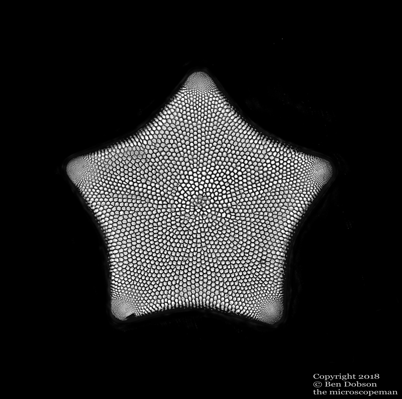
Tricertatium formosum A marine diatom 245 microns from 102 form slide by Klaus Kemp, using the immersion lens at x1000. Shot with a green filter which improves resolution slightly on my lens as it can’t bring yellow and blue light to a point. Apochromatic lenses that normally cost more 1000 each are required create perfect colour representation. It shows at high magnification. Sometimes I embrace this effect as can be seen in the different coloured (e.g. red) fringing Neon. Sometimes I use a filter and then switch to shades of grey.
I really love amazing geometric variation that diatoms display and certainly this five pointed star is a wonderful thing. I also like the texture here in black and white, like dry cracked earth.
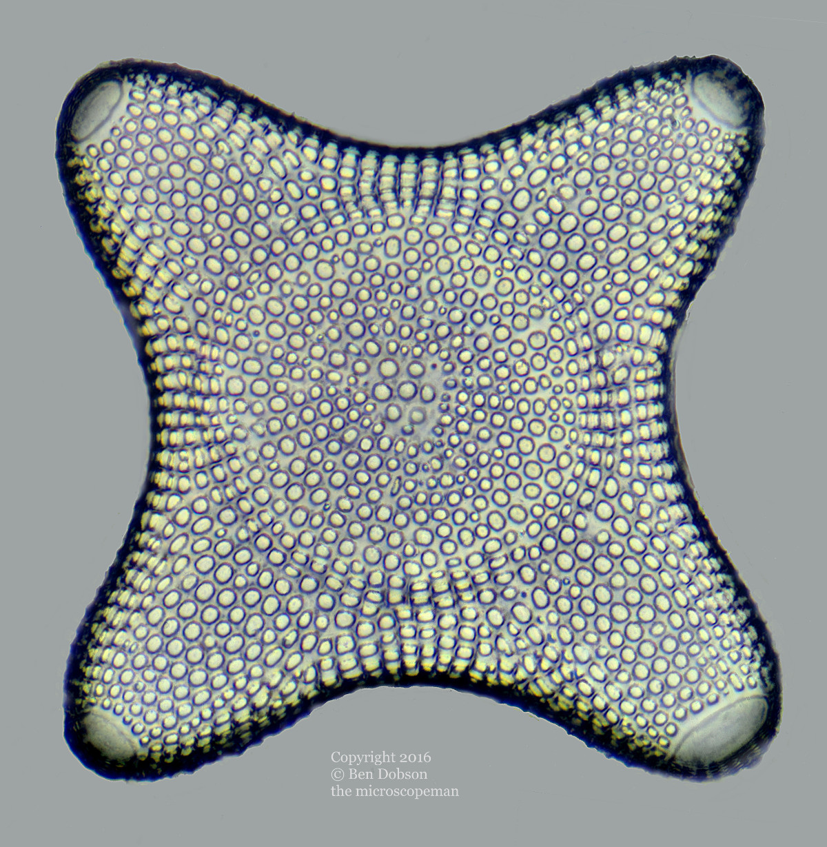
Triceratium antediluvianum
Another diatom 97 microns, at x 1000 again from Klaus’s slide.
Black Narcissus
 The image is of the base of the flower, the ovaries, of Narcissus cyclamineus sectioned at ten microns, 0.01mm , using an expensive professional microtome. The blades of the microtome are made by a Japanese company that still manufactures samurai swords.
Preparing some tissue for cutting that thin is quite time consuming and takes dozens of processing steps after fixing the tissue in formaldehyde , in this instance for a month. The tissue was stained with fast green and a red DNA stain called saffarinin. When viewed on one of my professional c-type prints it is possible to spot those cells that were undergoing cell division in the red stained ovaries when placed in formaldehyde. On smaller prints one must use a magnifying glass to see this, on a large print one needs to look closely.
The black background is digital and used to enhance the contrast in the image.
White Narcissus
Same slide, different rendering of image.

Astro Narcissus
Same slide, reshot using polarised Light

Moss leaf showing chloroplasts.
 The light harvesting sugar factories of the moss are the bright green circles, which are about four microns, or 0.004 mm. You can also see the cell walls.